

When Are Cleft Lips Repaired
Continuing Educational activity Activeness
Cleft lip repair is a surgical procedure that may exist approached with a wide array of dissimilar techniques. The ultimate goal of cleft lip repair is to restore the sphincter function of the orbicularis oris muscle and obtain a cosmetically favorable consequence for the developing child. This action reviews the almost usually used surgical techniques and highlights the importance of a multidisciplinary team in these complicated patients that include an otolaryngologist, a plastic surgeon, a dentist, an orthodontist, a pediatrician, a geneticist, a nurse, a social worker, and a speech-language pathologist.
Objectives:
- Discuss the goals and timing of crevice lip repair.
- Identify the surgical considerations for cleft lip repair.
- Describe the most common surgical techniques for unilateral and bilateral scissure lip repair.
Introduction
Crevice lip deformity is 1 of the nearly common congenital malformations.[i] It may be isolated, or it may be associated with other malformations. Management of the scissure lip patient is centered around a multidisciplinary approach. Ultimately, cleft lip requires surgical intervention for definitive repair for which many surgical techniques have been described. Preoperative and postoperative strategies assistance in the successful management of patients with this disorder. The choice of the surgical technique employed depends on many factors such equally surgeon preference, the severity of the deformity, and the presence or absence of other deformities. In this discussion, we aim to present the near usually used surgical techniques for cleft lip repair, their advantages, and their disadvantages.
Anatomy and Physiology
In club to appreciate the anatomy of cleft lip deformity, it is vital to have a thorough agreement of the embryology behind this disorder. The first pharyngeal arch begins to develop facial prominences effectually the 4th week of embryonic evolution. These structures are the maxillary prominences, derived from neural crest cells, and the nasal placodes, derived from surface ectoderm. Around the 5th week of embryonic development, the nasal placodes invaginate, forming the nasal pits, creating a ridge of tissue around the pits called the lateral nasal prominence at the lateral attribute and the medial nasal prominence at the medial aspect. During the sixth to seventh weeks of embryonic development, the bilateral maxillary prominences grow medially towards the medial nasal prominences, and fusion occurs, leading to the formation of the upper lip. The fusion of the bilateral medial nasal prominences forms the medial portion of the upper lip, whereas the paired maxillary prominences grade the lateral aspects of the upper lip. Further medial growth of the maxillary prominences leads to additional fusion of the deep medial nasal prominences, which creates the master palate. The principal palate includes the 4 incisors and the palate inductive to the incisive foramen. Additionally, the medial nasal prominences form the philtrum, the columella, and the nasal tip. The lateral nasal prominences are involved in the development of the olfactory organ and practise not contribute to upper lip germination.[two]
Disruption at any of the aforementioned points during upper lip embryologic development leads to cleft lip deformity, unilateral or bilateral. Cleft lip deformity tin can be further classified into complete and incomplete clefts based on the extent of the deformity. Complete crevice consists of disruption of the entire vertical thickness of the upper lip. It is normally associated with an alveolar defect since the alveolus is part of the primary palate. Incomplete fissure lip involves only a portion of the superlative of the upper lip and has a segment that is continuous between the two portions of the upper lip. The extent of incomplete scissure lip varies from muscular discontinuity with intact overlying skin to a broad cleft with a thin segment of skin that crosses the crevice, called the Simonart band.
Normally, the orbicularis oris muscle surrounds the lips to form a complete sphincter around the mouth. Patients with crack lip deformity have orbicularis oris deformities that must exist corrected to achieve proper lip and mouth office. Patients with microform crevice lip in which an incomplete crack involves less than two-thirds of the upper lip usually take an orbicularis oris muscle that connects the medial and lateral segments of the fissure at its upper segment. In contrast, the lower muscle segments insert into the scissure margins. Patients with complete cleft lip deformity accept an orbicularis oris muscle that fails to environs the mouth to form a sphincter. In these patients, muscle fibers run along the cleft margins to insert on the columella on the medial segment of the cleft and the nasal ala on the cleft's lateral segment. Correction of the scissure lip requires detachment of the orbicularis oris muscle'southward aberrant attachments and creating a horizontal bridge across the crevice to course a complete sphincter around the mouth. In bilateral cleft lip, the muscle fibers run along the lateral cleft margins to insert on the bilateral nasal alae. In these patients, the medial prolabial segment is commonly devoid of muscle tissue, which leads to anterior protrusion of the prolabial segment and main palate relative to the nasal septum.
When speaking about cleft lip deformity, it is important to also take into consideration the nasal deformity associated with this disorder. Nasal deformity associated with this disorder is caused by traction from the abnormally inserted orbicularis oris muscle and the absent inductive nasal flooring.[iii]
In unilateral crack lip, the lower lateral cartilage on the cleft side volition have a brusk medial crus and a long lateral crus in a more caudal position. The columella will be found on the noncleft side. These anatomical abnormalities lead to nasal tip difference towards the noncleft side with nasal septum deviation towards the noncleft side and a laterally, inferiorly, and posteriorly displaced alar base of operations on the cleft side.
In bilateral fissure lip, lack of orbicularis oris muscle in the prolabial segment causes inductive premaxillary protrusion. This causes the medial crura of the bilateral lower lateral cartilages to be abnormally short and the columellar skin to exist deficient, leading to a shortened, well-nigh absent, columella. These findings lead to a wide nasal tip with laterally displaced alae.
Indications
Any patient built-in with a cleft lip should undergo repair if no contraindications exist.
Contraindications
The following are some contraindications to cleft lip repair:
- Surgery may be delayed in case further workup of coexisting conditions is necessary.
- Weather that preclude toleration of general anesthesia may delay the surgery.
- Circumstantial conditions that require surgery on a more urgent basis, such as cardiovascular anomalies, should be performed prior to cleft lip repair.
Equipment
The procedure requires the post-obit equipment:
- Scalpel
- Ophthalmic scissors
- Tissue scissors
- Mosquito clamps
- Toothed pick-ups
- Non-toothed pick-ups
- Double-pronged peel hooks
- Ruler
- Sterile marker
Personnel
The post-obit personnel are needed:
- Surgeon
- Surgeon's banana
- Operating room technician
- Registered nurse
- Anesthesiologist
- Anesthetist
Preparation
Although some have proposed surgical correction of the cleft lip as early as 48 hours later birth, surgical repair is generally performed following the rule of 10s, which states that the timing should be performed at ten weeks of age, when the patient weighs 10 lbs. and when hemoglobin is at x g/dL.[4] Repair between 2 to iii months of historic period allows the patient to be adequately evaluated for other congenital anomalies and for the patient to abound, giving additional tissue for repair.
Before definitive surgical repair, techniques may be employed that facilitate surgical intervention, particularly for wide clefts. The almost widely used techniques are tape adhesion and orthopedic appliances.[5] The principal goal of presurgical techniques is the narrowing of the cleft to minimize lip tension later repair and ameliorate outcomes. Tape adhesion consists of the awarding of steri strips beyond the cleft to attain this goal. Orthopedic appliances, also termed nasoalveolar molding, consists of wearing a palatal appliance synthetic by an orthodontist. This palatal appliance brings the alveolar arches closer together and narrows the cleft. The palatal appliance is gradually changed over weeks to months to optimize cleft width for surgery.
Technique
The two most usually used surgical techniques for the repair of the unilateral cleft lip, and the ones that volition be discussed here, are the rotation-advancement technique, as well called the Millard technique, and the triangular flap technique, too known as the Tennison-Randall technique.[6][7]
The rotation-advancement technique was first described by Millard and consists of a rotational flap on the cleft's medial segment and an advancement flap originating from the cleft'due south lateral segment. The advantages of this technique include a suture line that recreates the philtrum on the scissure side; access to nasal tip cartilages assuasive nasal reconstruction; and allows intraoperative adjustment. Disadvantages have potential nostril stenosis, difficulty closing broad clefts, and increased difficulty for the inexperienced surgeon as the technique requires ample intraoperative judgment for flap modification as the surgery progresses.[8][9] The triangular flap technique was starting time described past Tennison and consists of a triangular flap originating from the cleft side inserted on an incision performed on the noncleft side. This technique's main advantage is that information technology'southward based on carefully measured and predetermined landmarks that allow the inexperienced surgeon to perform the procedure hands. Additionally, this flap allows wider clefts to be repaired. The disadvantages in this technique are generally related to cosmetic outcomes and include failure to create a philtrum on the cleft side, a scar that crosses cosmetic subunits, inability to access nasal cartilages during repair, and disability to change repair once surgery has started.[10]
Surgery will exist guided by important anatomical landmarks that should be identified before the procedure, regardless of technique (Figure one). These structures are marked with a marker. Structures that must be marked are: the noncleft side'due south Cupid's bow peak (i), the midpoint between both Cupid's bow peaks (two), the midpoint of the columella (3), the base of the columella on the noncleft side, and fissure side (4 and 5), nasal ala base on the noncleft side and cleft side (6 and 7), and the oral commissure on the noncleft side and cleft side (viii and 9). Later on these structures are identified, other landmarks necessary for proper surgical planning may be determined. The peak of Cupid's bow on the medial cleft side (10) is adamant by measuring the distance from the noncleft side's Cupid'south bow elevation to the midpoint betwixt both Cupid'southward bow peaks. This distance equals the distance between the midpoint betwixt both Cupid's bow peaks and the cleft side'due south Cupid's bow peak on the medial segment of the cleft. The Cupid's bow tiptop on the lateral cleft margin (ten') is determined by measuring the altitude from the noncleft side oral commissure to the noncleft side Cupid'southward bow and marking a point at the same altitude on the lateral crevice margin from the cleft side's oral commissure. These two points, the medial cleft margin Cupid's bow elevation and the lateral cleft margin Cupid'south bow elevation, are essential in creating continuity in the vermilion border during repair and are tattooed with a needle with gentian violet or methylene bluish before surgery.
After repair, the incisions are kept clean and free of crusting past cleaning with hydrogen peroxide and applying antibody ointment. Elbow splints are used for 3 weeks to prevent the patient from bending arms and touching surgical wounds. During the first postoperative week, arm restraints are also used. Feeding is done by a bulb syringe for 3 weeks. Sutures are removed afterward the first postoperative week. Antibiotic prophylaxis with a penicillin antibody is given during the first 5 postoperative days.
Millard Rotational-Advancement Technique
Before starting the procedure, the anatomical landmarks described above should be identified and marked. After identifying the proper anatomical landmarks, the incisional lines should be outlined (Figure 2). The start incisional line volition class the medial crack segment rotational flap. This line extends from the Cupid's bow peak on the medial scissure margin superiorly along the vermilion border and then curves medially towards the base of the columella (bespeak 10 to point 3). A second incisional line should be marked, which continues along the medial crevice margin's vermilion edge from the point the commencement incisional line leaves the vermilion edge and extended towards the nasal floor. This creates what is called a c-flap, which may exist utilized to extend the columella on the cleft side when required (Figure iii). The incision which will form the lateral advocacy flap should so be traced. This incision volition extend from the Cupid's bow peak on the cleft's lateral margin superiorly along the vermilion edge towards the nasal floor (From indicate ten to point 7). Another incision line is then traced on the lateral cleft margin from the junction of the ala with the lip towards the lateral alar margin, post-obit the alar margin horizontally and and so vertically.
After the proper anatomical landmarks are identified and the incisional lines traced, repair may exist performed equally follows:
- A curvilinear incision is fabricated from the scissure side'due south Cupid's bow peak on the medial crack margin towards the base of the columella. This incision should involve the pare only.
- An incision is fabricated from the cleft side'south Cupid's bow peak on the medial crack margin towards the nasal flooring along the vermilion border. This incision involves the pare and subcutaneous tissue, leaving the oral mucosa intact.
- An incision is performed on the lateral cleft margin extending from the cleft side's Cupid's bow height on the lateral cleft margin superiorly to the nasal floor along the vermilion border. This incision should be through and through involving the skin, subcutaneous tissue, and mucosa.
- The orbicularis oris muscle is exposed by undermining the skin from the underlying muscle 1 cm from the incision margins in both the medial and lateral cleft segments. Undermining of the oral mucosa from the overlying muscle must also exist done using the aforementioned distance from the incision margins.
- The orbicularis oris musculus's aberrant attachments to the base of operations of the columella on the medial cleft margin and the alar base on the lateral cleft margin are transected to create bilateral muscle flaps.
- The skin is dissected off of the columella, and dissection is directed in a cephalad direction between both medial crura of the bilateral lower nasal cartilages to gain admission to the nasal dome and elevate the skin off of the dome.
- The nasal ala on the cleft side is detached from the underlying maxilla, and the skin is elevated off of the lower cartilage's lateral crus on the cleft side. The nasal mucosa is also dissected off of the lower cartilage's lateral crus on the crack side.
- A skin claw is used for upwards retraction of the peel overlying the dome of the cleft side'southward lower nasal cartilage. At this moment, the medial cleft margin'due south c-flap may be sutured to the medial scissure incisional margin to elongate the columellar skin on the cleft side (Effigy 4).
- The medially-based rotational flap is rotated inferiorly, and the laterally-based advancement flap is extended medially. The closure is assessed to determine if additional rotation will be necessary to close the defect. If rotation is insufficient, a dorsum-cut may exist performed at a perpendicular angle inferiorly from the columellar base margin of the incision performed in step one.
- After acceptable closure of both flaps is confirmed to be possible, the bilateral muscle flaps created in step 5 may be rotated inferiorly and sutured together with interrupted sutures.
- A traction suture is placed betwixt the medial crack segment Cupid's bow peak and the lateral cleft segment Cupid's bow peak.
- The deep portion of the superior medial role of the lateral advancement flap is sutured to the anterior nasal spine. This suture anchors the lateral advocacy flap.
- The bilateral nasal mucosal flaps obtained from the extension of the incisions along the vermilion border into the anterior nasal floor are sutured with interrupted chromic sutures to shut the anterior nasal flooring. During this footstep, it is important to pay attention to the possible narrowing of the nostril on the cleft side. If this occurs, an elliptical incision may be performed at the lateral alar margin of skin only, and the pare removed—this aids in achieving more than anterior positioning of the retroplaced nasal ala.
- The peel is closed with interrupted nylon sutures (Effigy 5).
- The vermilion and the vermilion border are so closed with interrupted nylon sutures on the outer surface and with interrupted chromic sutures on the inner surface.
- The oral mucosa is closed with interrupted chromic sutures.
- A eternalize is placed at the cleft side's nostril to stabilize the repaired anterior nasal floor and avoid nostril stenosis.
Tennison-Randal Triangular Flap Technique
Prior to commencement the process, the same anatomical landmarks described for the Millard technique should be identified and marked. Additionally, surgical planning must be washed with farthermost care since this technique allows for little intraoperative modification. Surgical planning starts with identifying the length the lip must cover. This is done past calculating the deviation betwixt the base of operations of the columella and Cupid's bow summit on the noncleft side and the base of the columella and Cupid'due south bow top on the crevice side'south medial margin (Effigy vi). The result of this difference is the number of millimeters the lip must exist lengthened [(A to B) – (C to D) = Cleft width]. To account for scar contracture, 2 mm should also be added to this outcome. When this altitude is greater than 6 mm, a two-triangular flap technique should be employed with a laterally-based inferior triangular flap and a medially-based superior triangular flap.[eleven] The incisions to be performed are then outlined, starting with the laterally-based triangular flap (Effigy vii). This incision line starts from the Cupid's bow peak on the lateral margin of the fissure and extends superiorly along the vermilion border the length that the lip must be lengthened up to a maximum of 6 mm. A line that is the aforementioned length every bit the first line is and then drawn perpendicular to the vermilion border. The distance from point A to point B equals the distance from bespeak B to indicate C, which in turn equals the distance from signal C to betoken A, creating an equilateral triangle flap. An incisional line that is the aforementioned length every bit these lines are and then drawn perpendicularly from Cupid'south bow acme on the fissure side'due south medial margin. This is where the laterally-based triangular flap will be inserted. If a two-triangular flap technique is to exist employed, the superior, medially-based triangular flap is traced starting with a line perpendicular to the superior medial cleft margin vermilion border that will exist directed towards the base of the columella, and its length will be the additional corporeality of millimeters required across the initial 6 mm. The other side of the equilateral triangle is traced from the bespeak this line meets the vermilion border superiorly towards the nasal floor. The contralateral incision line where this triangle will exist inserted is traced perpendicularly from the vermilion border at the junction of the alar border and the upper lip.
After the surgical incisions are properly measured and outlined, the surgery may brainstorm using the following steps:
- An incision is performed on the medial cleft margin from the medial scissure Cupid's bow superlative to the anterior nasal flooring along the vermilion edge. This incision is made through the peel and subcutaneous tissue; the oral mucosa is left intact (Figure 8).
- An incision is made from the medial cleft of Cupid's bow peak to betoken B'. This incision is through the pare just. If a second triangular flap is to be used, the incision on the medial cleft margin from the vermilion edge to point E is performed in this step as well and is through the skin but.
- On the lateral crevice margin, a through-and-through incision is performed from the lateral cleft Cupid'due south bow pinnacle superiorly to the anterior nasal floor along the vermilion border.
- An incision is made from point B to point C that is through the skin simply.
- A curvilinear incision extended superomedially from point C to join the vermilion edge is performed and is through the skin only. This will create a triangular piece of skin medial to the incision that is discarded.
- The orbicularis oris musculus is dissected free from the overlying peel and underlying oral mucosa on both crack margins, a distance of 1 cm from the margins.
- The attachments of the orbicularis oris muscle to the base of the columella and the alar base of operations are transected.
- The alar base on the cleft side may be dissected from the maxilla if repositioning of the ala is desired.
- The nasal mucosa is dissected free from the septum and from the cleft side'due south lower lateral nasal wall to create the flaps necessary to develop the anterior nasal flooring. These flaps are and then sutured together with interrupted chromic sutures.
- The scissure side'due south nasal ala may be sutured to the anterior nasal spine with a polyglactin suture and tightened until the ala reaches the desired position.
- The bilateral orbicularis oris muscle flaps created in stride 7 are rotated inferiorly and sutured with interrupted polyglactin sutures to repair the muscular sphincter.
- A nylon suture is used to estimate the medial and lateral crack of Cupid'south bow peaks.
- The triangular flaps are inserted into the proper positions, and a nylon corner suture is used between points B and B' and points D and D' if a second triangular flap was used.
- The upper lip pare is and then sutured with interrupted nylon sutures.
- Interrupted nylon sutures are used to approximate the external surface of the vermilion.
- Interrupted chromic sutures are used to approximate the internal surface of the vermilion and the oral mucosa (Figure 9).
Bilateral Fissure Lip Repair
Bilateral cleft lip repair poses additional difficulty when compared to unilateral cleft lip repair. Different surgeons have different techniques ranging from 2-stage surgical repairs to single-stage surgical repair. Some surgeons utilise the triangular flap technique as a single-stage or a 2-phase procedure. Here, we will discuss the single-stage repair introduced by Millard.[12][thirteen]
The almost hard part of bilateral cleft lip repair is managing the premaxilla, which may be anteriorly and sagitally displaced to either side as a result of it being fastened only to the nasal septum, and the prolabium, which may be as well small to provide sufficient tissue for cleft repair. These challenges can exist handled by unlike methods, which include performing surgical lip adhesion before definitive surgical repair of bilateral crack lip, using presurgical orthopedic prostheses or surgical procedures to bring the premaxilla to a more than inferior and posterior position.
Every bit with unilateral cleft lip repair, identifying anatomical landmarks and outlining surgical incisions are important prior to first surgery (Effigy 10). First, the midline of the prolabium is identified (point 1). Two symmetrical incision lines are traced in the prolabium, these incisions begin only medial to the base of operations of the columella and continue inferiorly until they reach the vermilion edge, diverging laterally a small amount. The upper lip's vertical top will be adamant by the length of these incision lines on the prolabium, not by the lateral lip segments. If a marked discrepancy exists, the height of the lateral lip segments may be adjusted by the excision of crescent-shaped segments of peel nether the alar bases on the lateral lip segments. Incision lines on the lateral lip segments are traced beginning at both alar bases and extending inferiorly along the vermilion border a distance equal to the prolabium incisional lines (line ii-iii equals line 4-5, which equals line half dozen-7, which equals line 8-ix). Incisional lines are drawn beyond the bilateral lip segment vermilion that will create flaps D and E that will comprise vermilion, mucosa, submucosa, and orbicularis oris muscle. The lateral alar bases are marked to trace incision lines forth the alar bases. After outlining incision lines, surgery may be performed using the following steps:
- Incisions two-3 and 4-5 are performed on the prolabium, creating the prolabial flap (A) and bilateral forked flaps (B and C).
- On the prolabium, incisions may be extended inferiorly into the vermilion, and a horizontal incision may exist performed across the vermilion border (three-5) to create flap H that may be used to add book to the vermilion if whistle deformity occurs.
- Incisions 6-vii and eight-9 are performed on the lateral lip segments along the vermilion border.
- Flaps D and E are created on the lateral lip segments by incisions across the vermilion.
- Circumalar incisions half-dozen-12 and 8-xiii are performed. The lateral lip segments are dissected from the maxilla to allow medial advancement. The nasal alae are freed from the maxilla to allow medial advocacy.
- The tips of the nasal ala are brought medially and sutured to the area of the anterior nasal spine to attain symmetric nostrils.
- Excess vermilion in the prolabium is excised and sutured to the premaxilla to provide midline upper lip volume.
- The upper edges of the lateral lip flaps (F and G) are brought to the midline and sutured to the area of the inductive nasal spine inferior to where the nasal alae were sutured. These flaps are not sutured to one another to allow space for the prolabial flap A.
- Orbicularis oris muscle found in flaps D and E is brought midline and sutured to create the muscular sphincter.
- The prolabial flap is lowered, and a suture is placed from its undersurface to the orbicularis oris muscle to create a philtral dimple.
- The bilateral forked flaps B and C are sutured to the lateral anterior nasal walls to create the bilateral inductive nasal floors.
- The skin and mucosa are sutured to finish the repair.
- If a central vermilion deficiency occurs leading to whistle deformity, flap H may be tucked between the prolabial flap A and the bilateral vermilion flaps D and E to add volume to the central vermilion.[xiv]
Complications
The complications related to surgical repair are wound dehiscence, scar contracture, scar hypertrophy, and infection. Other complications are related to lip and nasal deformities not resolved during the primary repair, such as vermillion notching, misalignment of the white gyre, orbicularis discontinuity, short or excessive length of the lip, brusque or deviated columella, the horizontal orientation of the nares, abnormalities of nostril size, and disturbance of alar base position.
Avoiding tension at closure and postoperative local wound care will subtract the incidence of wound dehiscence. Careful marking and approximation of the white curlicue are necessary to avoid its misalignment. Conscientious orbicularis muscle autopsy and suturing are also necessary to avoid orbicularis muscle aperture. Prevention is as always therefore the best class of activeness to avoid complications.
Enhancing Healthcare Team Outcomes
The standard of care for a patient with cleft lip involves a multidisciplinary team that meets multiple times each yr to hash out patient management. This squad should be comprised of an otolaryngologist, a plastic surgeon, an oral and maxillofacial surgeon, a speech-linguistic communication pathologist, an orthodontist, a dentist, a pediatrician, a geneticist, an audiologist, nurses, social workers, and a psychologist. Each member of the team provides their expertise to optimize the management of patients that will most likely take disorders associated or due to their crevice lip deformity.[15][xvi]
(Click Image to Enlarge)
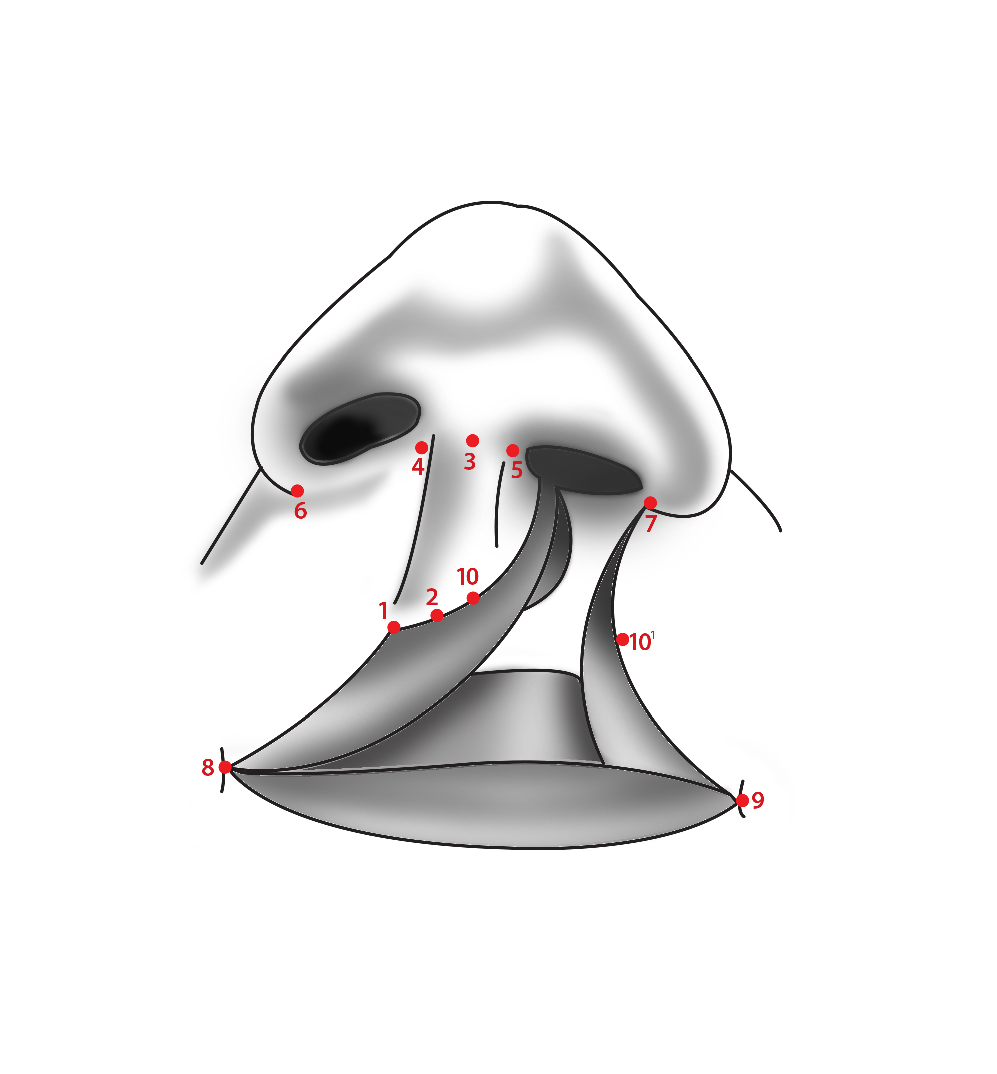
Effigy one. Fissure lip anatomic landmarks.
Drawing by Antonio Riera, Medico. Digitalization by Amarilys Irizarry.
(Click Image to Enlarge)
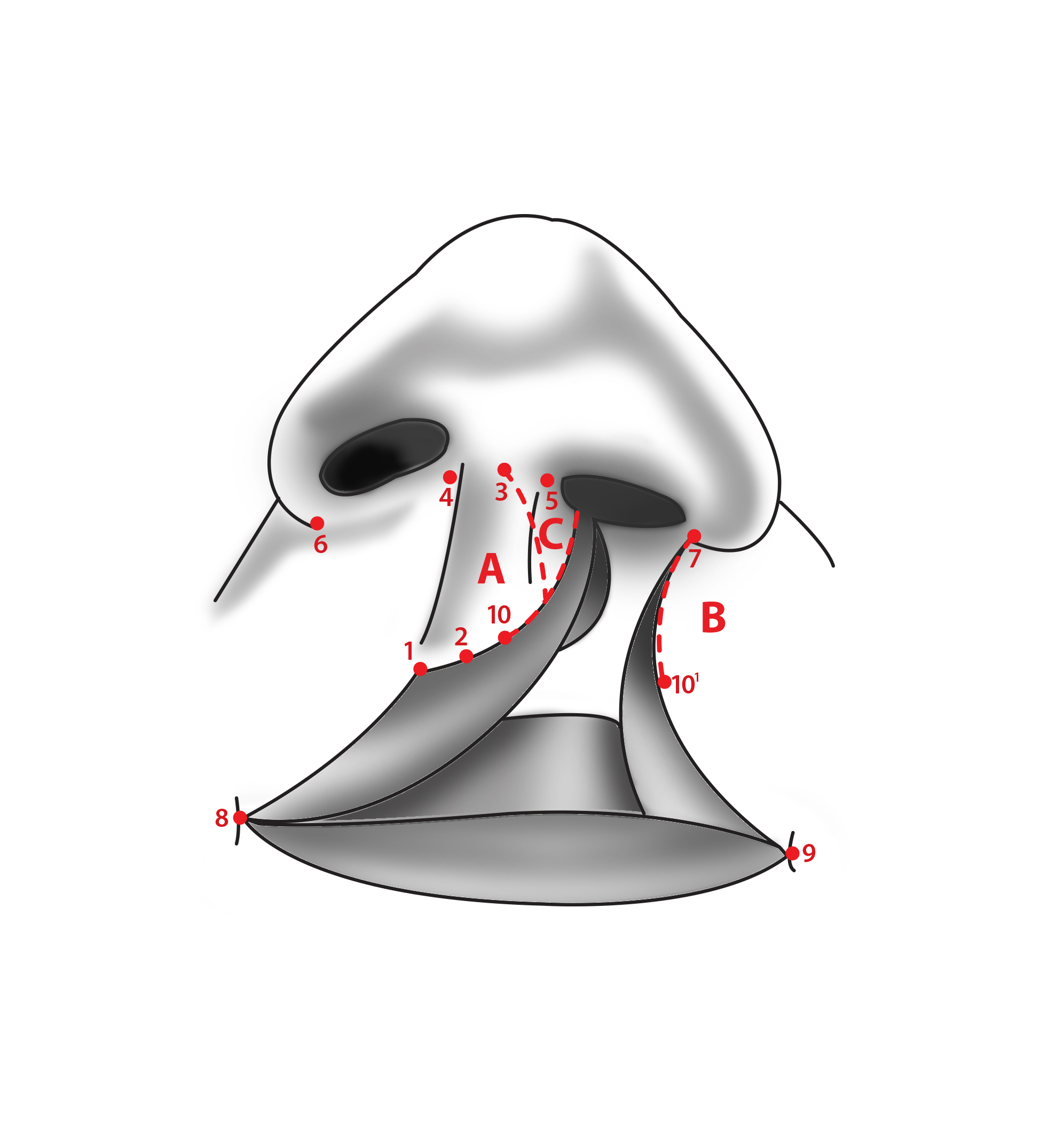
Effigy 2. Millard rotation-advancement technique incision lines.
Drawing by Antonio Riera, MD. Digitalization past Amarilys Irizarry.
(Click Image to Enlarge)
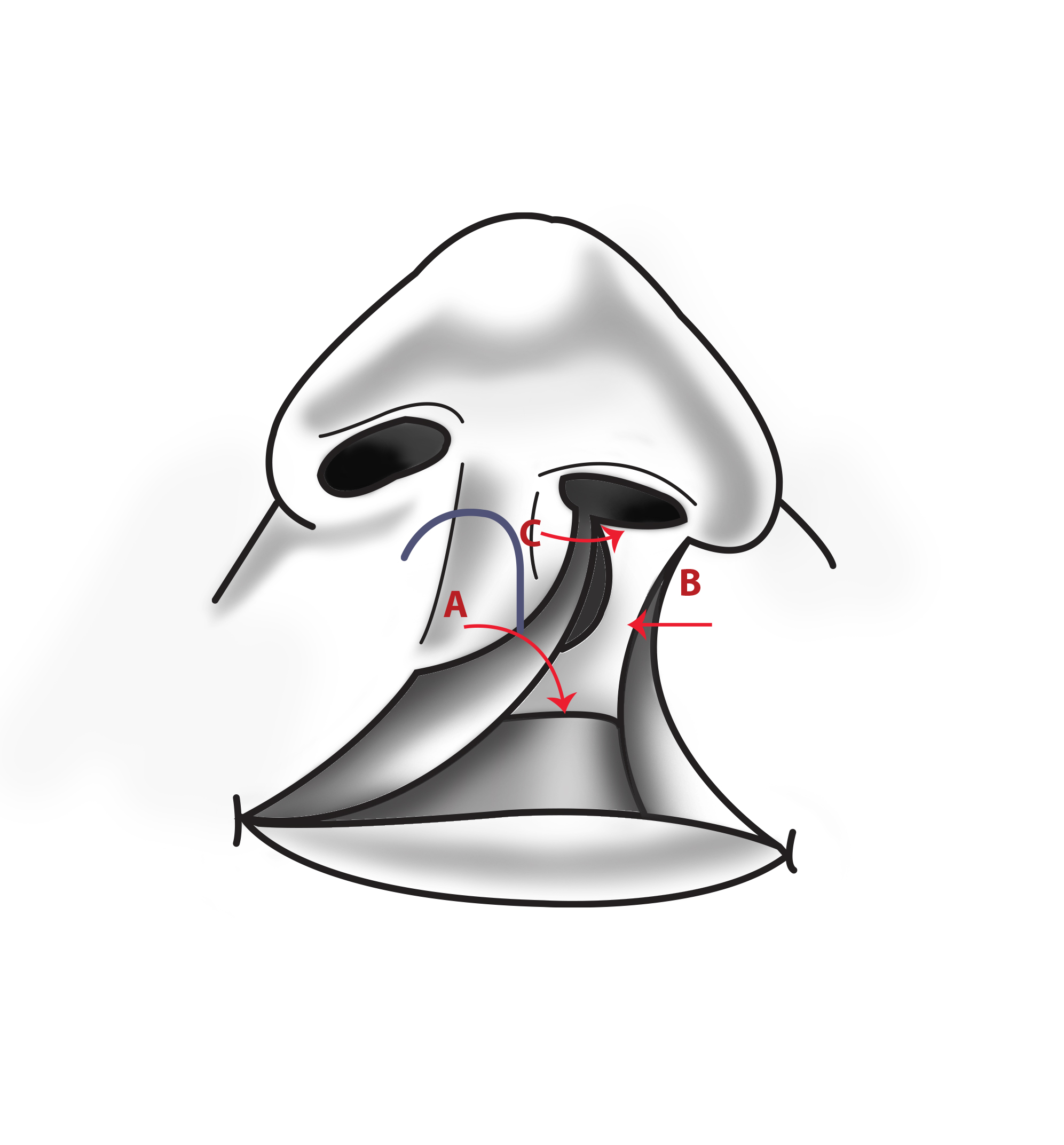
Effigy 3. Millard rotation-advancement technique flaps.
Drawing by Antonio Riera, MD. Digitalization by Amarilys Irizarry.
(Click Image to Enlarge)
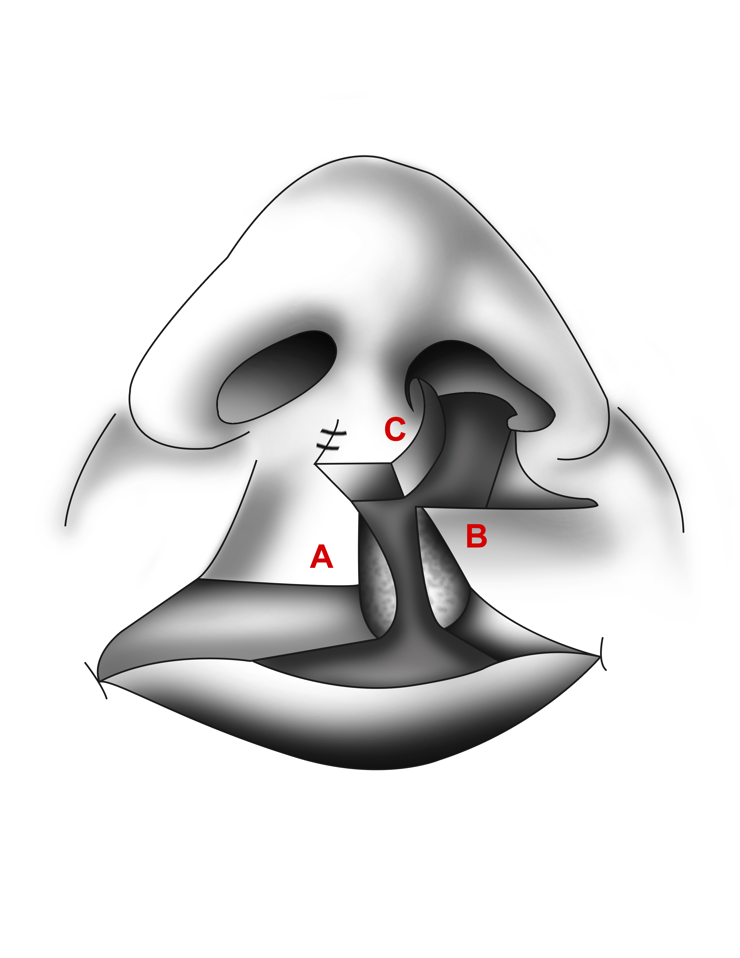
Figure 4. Millard rotation-advocacy technique with c-flap sutured for columellar lengthening.
Drawing by Antonio Riera, Md. Digitalization past Amarilys Irizarry.
(Click Image to Overstate)
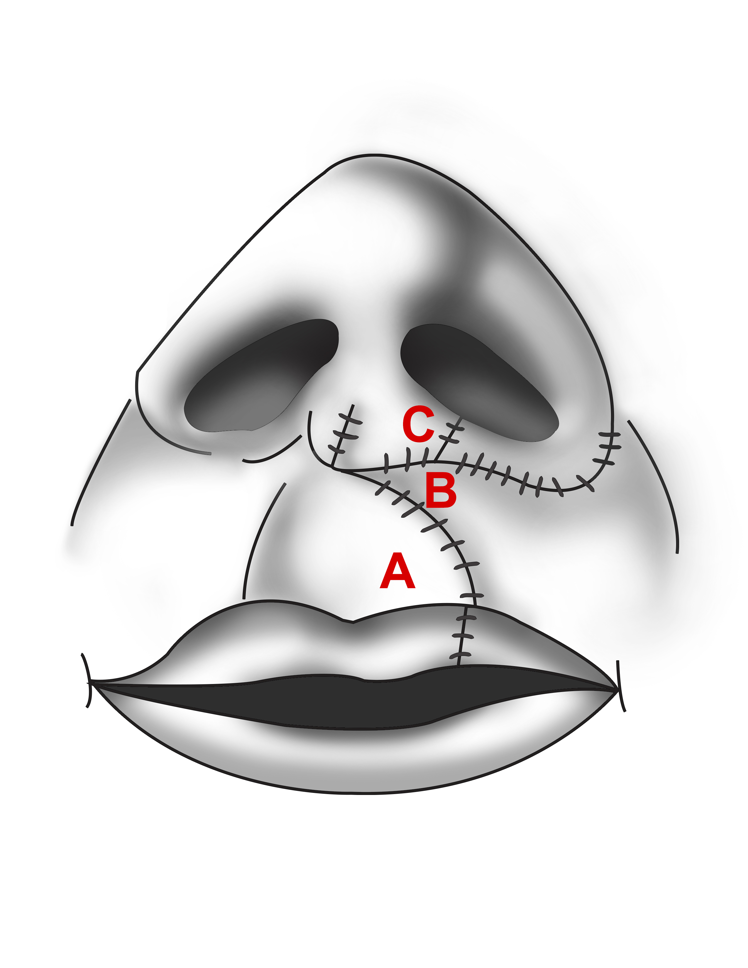
Figure v. Millard rotation-advancement technique.
Drawing by Antonio Riera, MD. Digitalization by Amarilys Irizarry.
References
[1]
Gatti GL,Freda Northward,Giacomina A,Montemagni G,Sisti A, Cleft Lip and Palate Repair. The Journal of craniofacial surgery. 2017 Nov; [PubMed PMID: 29088690]
[two]
Smarius B,Loozen C,Manten W,Bekker M,Pistorius Fifty,Breugem C, Accurate diagnosis of prenatal cleft lip/palate by understanding the embryology. Globe journal of methodology. 2017 Sep 26; [PubMed PMID: 29026689]
[iv]
Burianova I,Kulihova M,Vitkova V,Janota J, Breastfeeding Later Early Repair of Scissure Lip in Newborns With Cleft Lip or Crevice Lip and Palate in a Babe-Friendly Designated Hospital. Journal of man lactation : official periodical of International Lactation Consultant Association. 2017 Aug; [PubMed PMID: 28604150]
[v]
Thierens L,Brusselaers Due north,De Roo N,De Pauw G, Furnishings of labial adhesion on maxillary arch dimensions and nasolabial esthetics in cleft lip and palate: a systematic review. Oral diseases. 2017 Oct; [PubMed PMID: 27878905]
[six]
Han Yard,Park J,Lee Due south,Jeong W, Personal technique for definite repair of complete unilateral cleft lip: modified Millard technique. Athenaeum of craniofacial surgery. 2018 Mar; [PubMed PMID: 29609427]
[seven]
Rossell-Perry P, A 20-twelvemonth experience in unilateral cleft lip repair: From Millard to the triple unilimb Z-plasty technique. Indian journal of plastic surgery : official publication of the Association of Plastic Surgeons of India. 2016 Sep-Dec; [PubMed PMID: 28216814]
[eight]
Roussel LO,Myers RP,Girotto JA, The Millard Rotation-Advancement Cleft Lip Repair: l Years of Modification. The Crevice palate-craniofacial journal : official publication of the American Crack Palate-Craniofacial Association. 2015 November; [PubMed PMID: 25642967]
[nine]
Chang LS,Son Y,Baek RM,Kim BK, Anatomical Reconstruction of the Nasal Flooring in Complete Unilateral Cleft Lip Repair. Annals of plastic surgery. 2017 Oct; [PubMed PMID: 28570455]
[10]
Madaree A, Symmetric Philtral Column Repair for Unilateral Scissure Lip. Plastic and reconstructive surgery. 2019 Apr; [PubMed PMID: 30921134]
[xi]
Aranmolate S,Aranmolate SO,Zeri RS,Gbeneol T,Ajani AO, Upper Triangular Flap in Unilateral Cleft Lip Repair. The Journal of craniofacial surgery. 2016 May; [PubMed PMID: 26982113]
[12]
Trier WC, Repair of bilateral cleft lip: Millard's technique. Clinics in plastic surgery. 1985 Oct; [PubMed PMID: 3905170]
[13]
Adeyemo WL,James O,Adeyemi MO,Ogunlewe MO,Ladeinde AL,Butali A,Taiwo OA,Emeka CI,Ayodele AO,Ugwumba CU, An evaluation of surgical outcome of bilateral cleft lip surgery using a modified Millard's (Fork Flap) technique. African journal of paediatric surgery : AJPS. 2013 October-Dec; [PubMed PMID: 24469478]
[14]
Rossell-Perry P,Gavino-Gutierrez AM, Surgical Technique for Whistler Deformity Repair in Bilateral Scissure Lip Patients: An Anthropometric Study. Annals of plastic surgery. 2016 Aug; [PubMed PMID: 25057916]
[15]
Sánchez-Ruiz I,González Landa Grand,Pérez González Five,Díez Rodríguez R,López-Cedrún JL,Miró Viar J,García Miñaur S,de Celis Vara R,Sánchez Fernández L, [Integrated treatment of cleft lip and palate. Organisation of a treatment team]. Cirugia pediatrica : organo oficial de la Sociedad Espanola de Cirugia Pediatrica. 1999 Jan; [PubMed PMID: 10198542]
[16]
Fan KL,Black CK,Mantilla-Rivas Eastward,Bulas DI,Rubio E,Blask AR,Robinson C,Oh AK, Coordination of the Fetal Medicine Plant and the Cleft and Craniofacial Center: Awarding to Early Management of Infants With Crack Lip and Palate. The Periodical of craniofacial surgery. 2019 Oct; [PubMed PMID: 31524754]

Source: https://www.statpearls.com/ArticleLibrary/viewarticle/19598
Posted by: snowfamere.blogspot.com
0 Response to "When Are Cleft Lips Repaired"
Post a Comment